Highlights
The 3D Accuitomo 170 offers unsurpassed image clarity. With 9 fields of view and multiple acquisition modes, the 3D
Accuitomo 170 can meet all of your diagnostic needs with unparalleled quality. Its super-fine minimal voxel size of just 80
µm allows diagnosing even the most subtle details of bone and dentition. The 3D Accuitomo 170 is highly recommended by leading radiologists for periodontology, oral surgery, endodontics, orthodontics, dental implants, for the maxillofacial region and beyond.
- High – Quality Images
With Low Dose - Nine Fields Of View
- Four Imaging Modes
- Simple, Accurate
Positioning - Sharing Image Data
- Numerous Clinical Cases
Features
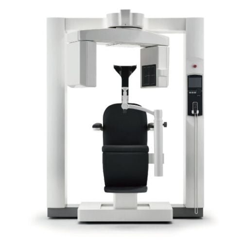
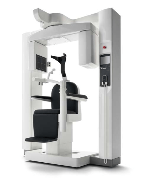
Unsurpassed Image Clarity
The 3D Accuitomo 170 offers unsurpassed image clarity. With 9 fields of view and multiple acquisition modes, the 3D Accuitomo 170 can meet all of your diagnostic needs with unparalleled quality. Its super-fine minimal voxel size of just 80 μm allows diagnosing even the most subtle details of bone and dentition. The 3D Accuitomo 170 is highly recommended by leading dental radiologists for periodontology, oral surgery, endodontics, orthodontics, dental implants, for the maxillofacial region and beyond.
Stunning Clarity
The high resolution 80μm acquisitions provide images of stunning clarity, giving you diagnostic information like you’ve never seen before. Take your treatment planning to the next level.
Nine Field of View
9 different fields of view can be selected to meet a wide variety of clinical needs. From the smallest, 40×40 to the largest 170×120, there is a size that fits your needs while always ensuring the lowest X-ray doses possible to the patient.
- Ø 170 x 120 mm
- Ø 170 x 50 mm
- Ø 140 x 100 mm
- Ø 140 x 50 mm
- Ø 100 x 100 mm
- Ø 100 x 50 mm
- Ø 80 x 80 mm
- Ø 60 x 60 mm
- Ø 40 x 40 mm
This flexibility allows the 3D Accuitomo 170 to provide stunning images for Endodontics, Periodontics, Otolaryngology, Maxillofacial surgery and many more. Resolution stays high and distortion is minimized for all regions.
Adaptable Acquisition Modes –
High Resolution Mode (Hi-Res)
At 1/4 the standard pixel size, high-resolution mode produces the
sharpest and clearest images the 3D Accuitomo 170 has to offer.
Even in hi-res mode, 360 scans take only 30.8 seconds, and 180
scans a mere 15.8 seconds. Available for 40 x 40 mm and 60 x 60
mm FOVs, it is ideal for observation of delicate bone structures such as the ossicular chain.
High Fidelity Mode (Hi-Fi)
Slow and steady scans at 30.8s for 360 degrees and 15.8s for 180
makes for exceptionally clear images with minimal artifacts. Zoom
reconstructions made from this acquisition are exceptionally clear.
High Speed Mode (Hi-Speed)
Full scan: 10.5s. Half scan: 5.4s. Reduces motion artifacts for patients that may not be able to sit still such as children and for patients concerned with higher X-ray dosage. Available for 40 x 40mm and 60 x 60 mm FOVs.
Standard Mode (Std)
Standard mode offers images of exceptional clarity and is suitable
for limited and wide views of temporal bone, paranasal, sinus,
maxilla and mandible, individual teeth etc.
Simple, Accurate Positioning
The three positioning laser beams and an LCD make patient positioning easy. The chinrest stabilizes the patient`s head to avoid movement. Scout images enable even more accurate positioning.
Two – Direction Scout
For even more accurate positioning, scout images can be utilized. After positioning, two still X-ray images of coronal and sagittal views can be taken to confirm that the position is accurate. If adjustment is necessary, positioning can be changed by dragging the cursor on the monitor and moving it to the center of the region of interest.
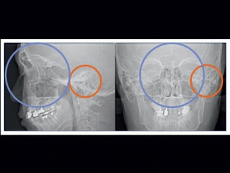
Use scout to accurately determine the minimal region of interest before exposing the patient to the higher dosage CT scan.
Easy as One,Two,Three
First, the patient`s initial position is set and recorded using the three positioning laser beams. Then, the region of interest is aligned in the LCD. The chair automatically moves into the optimal position. During the X-ray exposure, the patient is stabilized by the chinrest and the
headrest.
Acquisition to Diagnosis Made Simple
The i-Dixel imaging software offers a wide variety of features to help you quickly and easily create comprehensive treatment plans and explain those plans to your patients.
Freedom from Platform and Simplicity of Design
i-Dixel WEB runs as a web service on an X-ray server PC included with your Morita X-ray system. It serves as a local and secure web-based dental image processing service that you can access throughout your practice on a wide range of devices from workstations to tablets.
No Software Installation Needed
With the latest advancements in web technology, i-Dixel WEB gives youthe freedom to view your images wherever you want and however you want. Gone are the days of complicated chairside PC setups, and limited choices of hardware. Mac OS X and even iPads can be used to view and edit data from a Morita X-ray system.
One Volume Viewer Software
Share your cases with referring doctors or view them on computers without i-Dixel installed using the Morita One Volume Viewer.
CT data can be exported from i-Dixel or even i-Dixel WEB and copied to a DVD or thumbdrive and shared. What you see is what you get and any indication, annotations or even your contrast settings are visible no matter where the data is viewed.
New annotations can be added and contrast adjusted to suite the viewing environment but your indications are always visible and cannot be changed. What you can do with the 3D viewer in i-Dixel, you can do with One Volume Viewer too.
i-Dixel Conforms to the Following DICOM Standards:
- Modality Worklist Management Service Class (Optional)
- Storage Service Class
- Modality Performed Procedure Step Service Class
- Print Management Service Class
For more information on i-Dixel, please visit the i-Dixel webpage. To see the latest advancements in web technology and how this can simplify your IT needs, visit our i-Dixel WEB page for details.
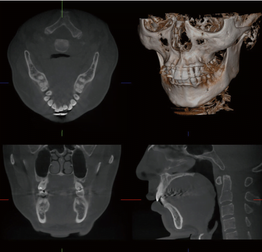
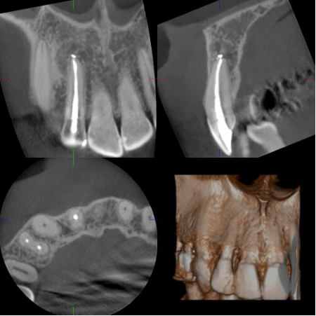
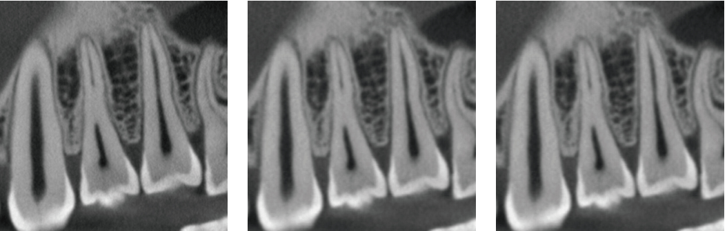
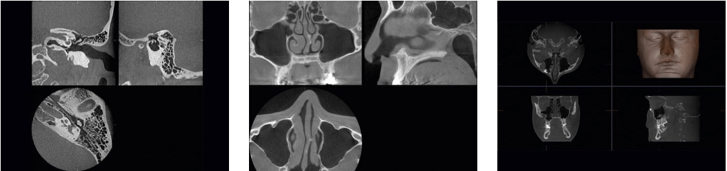
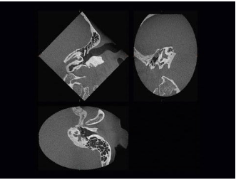
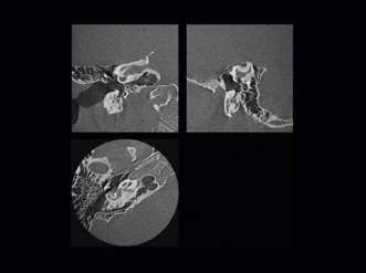
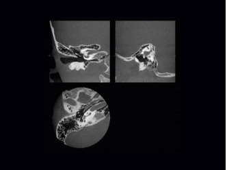
Clinical Cases
Doctors and Radiologists from all over the world have shared with us stunning cases that highlight the benefits of the 3D Accuitomo’s superior image quality in treatment planing and diagnostics.
Technical Detail
Spesifications
Nama : 3D Accuitomo
XYZ Slice View Tomograph
Model : MCT – 1
Type : EX1/2 F17
Power Supply : AC 100/110/120V
AC 220/230/240 vac
Power Consumtion : max 2.0 kVa
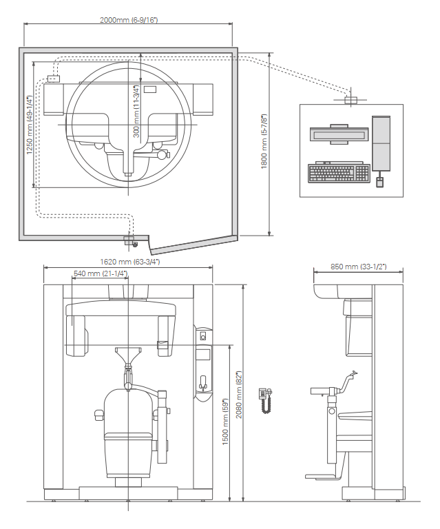
Main Unit : W1,620 mm x D1,250 mm x H2,080 mm
(63-3/4” x 49-1/4” x 82”)
Control Box : W100 mm x D40 mm x H115 mm
(4” x 1-5/8” x 4-1/2”)
Weight : Approx. 400kg (Approx. 882lbs)
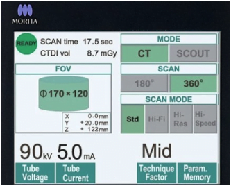
X-Ray Tube Voltage : 60-90 kV
X-Ray Tube Curnet : 1-10 mA
(Max 8mA : Hi-Fi, Hi-Res Mode)
Focal Spot Size : 0,5
Exposure Time : Std Mode: 17.5 / 9.0 sec
(360º/180º) Hi-Fi Mode : 30.8 / 15.8 sec
Hi-Res Mode : 30.8 / 15.8 sec
Hi-Speed Mode : 10.5 / 5.4 sec
Field of View : ø 40 x H40 mm
ø 60 x H60 mm
ø 80 x H80 mm
ø 100 x H50 mm
ø 100 x H100 mm
ø 140 x H50 mm
ø 140 x H100 mm
ø 170 x H120 mm
Voxel Size : 80 µm / 125 µm / 160 µm / 250 µm
* X-ray protection should be provided for the patient when X-rays are emitted.
* Design and specifications are subject to change without notification
Dimensions